
Background
Every MRI system makes use of radio-frequency (RF) fields. RF coils are used to generate RF fields to excite the atomic nuclei (transmit). The same or other RF coils are used to detect the RF signals from the atomic nuclei when these relax to their ground state (receive). The interactions of the RF coils, RF fields and the patient tissue are governed by the fundamental laws of electromagnetics and have a strong impact on the image quality. Particularly for ultrahigh field systems, the development of novel RF coils and antennas is an active field of research. The RF fields also pose challenging safety concerns, for example through heating of tissue or even more challenging, through heating of conductive implants in patients with medical implants. In the Electromagnetics in MRI group, we are trying to solve these electromagnetic puzzles by employing our arsenal of simulation and experimental methods.
RF antenna development
RF coil design heavily determines the image quality of MRI. Especially at ultra-high field strength (7T and beyond), coil design becomes of increasing importance for image quality and also for safety. Our group has a long standing track record of innovative RF coil designs for ultrahigh field (body) imaging, aiming for maximum efficiency, homogeneity and ease-of-use while minimizing power deposition within the patient tissue. We have made several key contributions to the introduction of the dipole antenna for ultrahigh field MRI. Our antenna designs have been adopted by various research sites across the world. Furthermore, our group is involved in the mCube consortium (http://www.mcube-project.eu/project/), where we aim to improve MRI by integrating metamaterials in RF antennas and by employing innovative coil designs such as coaxial cable antennas.
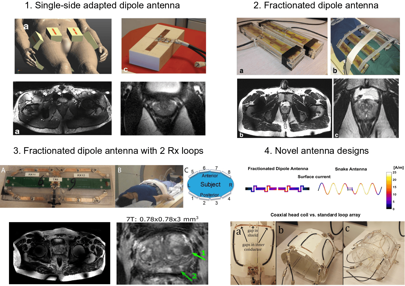
Figure 1: various dipole antenna designs (1-3) and prostate MRI images acquired with the dipole antennas at 7T. By changing the antenna design, image quality markedly improves. 4. Shows two novel antenna designs that have more recently been developed.
Relevant publications
- Raaijmakers AJE, Ipek O, Klomp DWJ, et al. Design of a radiative surface coil array element at 7 T: The single-side adapted dipole antenna. Magn Reson Med. 2011;66(5):1488-1497. https://doi.org/10.1002/mrm.22886;
- Raaijmakers AJE, Italiaander M, Voogt IJ, et al. The fractionated dipole antenna: A new antenna for body imaging at 7 Tesla. Magn Reson Med. 2016;75(3):1366-1374. https://doi.org/10.1002/mrm.25596;
- Steensma B, van de Moortele PF, Ertürk A, et al. Introduction of the snake antenna array: Geometry optimization of a sinusoidal dipole antenna for 10.5T body imaging with lower peak SAR. Magn Reson Med. 2020. https://doi:10.1002/mrm.28297
- Mollaei MSM, Van Leeuwen CC, Raaijmakers AJE, Simovski CR. Analysis of High Impedance Coils Both in Transmission and Reception Regimes. IEEE Access. 2020. https://doi.org/10.1109/ACCESS.2020.3009367
SAR and thermal safety of MRI
RF fields to not only generate signal. They also deposit heat which may cause tissue heating. Power deposition is expressed by the specific absorption rate (SAR). For safe operation of MRI systems, input power levels are constrained to predefined maximum levels. For high performance MRI systems operating at high field and/or with multiple RF transmit channels, power constraints are determined by limiting local temperature rise or the peak value in the corresponding SAR distributions. Since it is currently impossible to accurately measure temperature rise or SAR during MRI, constraints are typically determined by numerical electromagnetic simulations. Also, by using these simulations, we aim to improve our understanding and control over RF coils and RF heating in MRI. For this purpose, we use the software package Sim4Life of our collaboration partner Zurich Med Tech. We have developed various approaches to deal with local SAR and the corresponding uncertainties in multi-RF channel systems.In one recent innovative example, we have developed a deep learning method that can predict SAR distributions in patients undergoing an MRI exam based on the RF transmit field, which can be measured by the MRI scanner.
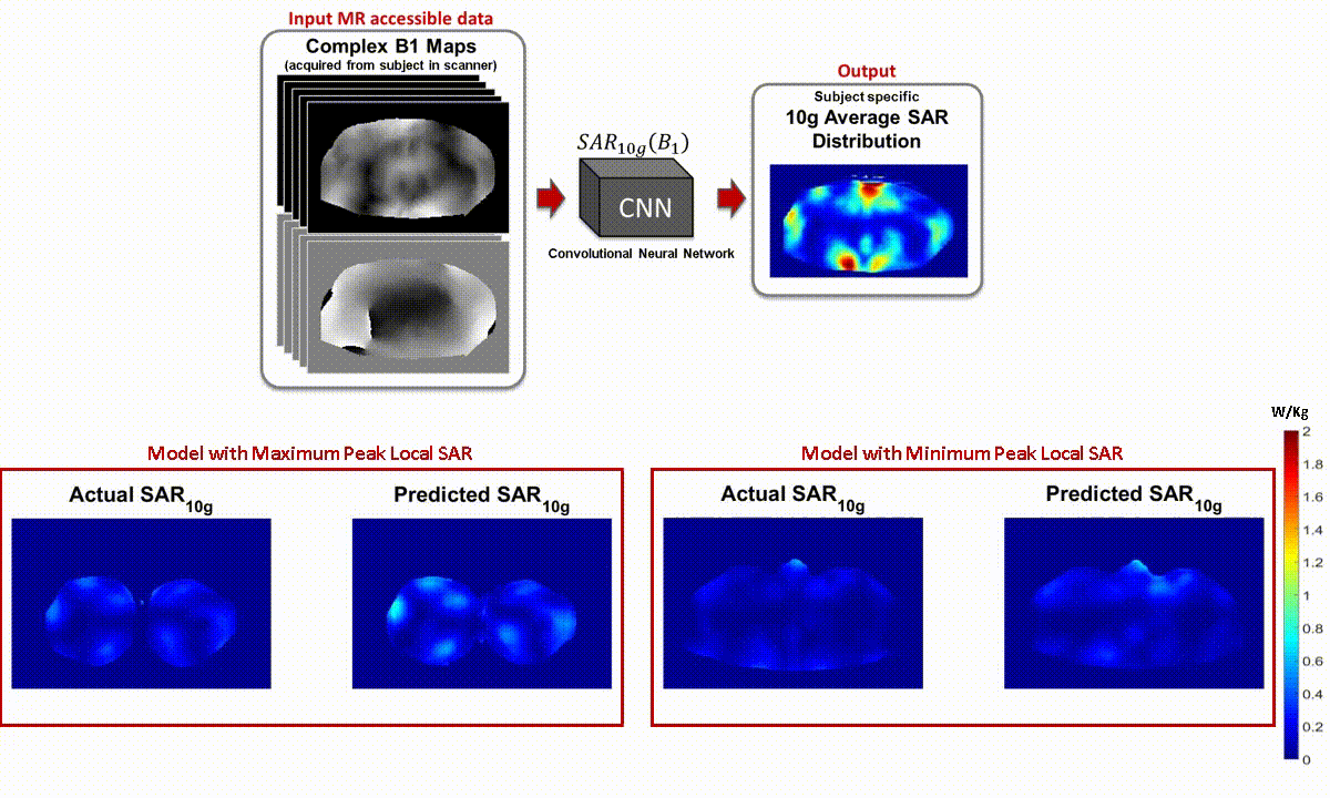
Figure 2: (top) deep learning method to predict local SAR from B1 maps measured on scanner. (bottom) comparison between ground truth simulated SAR, and SAR predicted from B1 map.
Relevant publications
- Simonis FFJ, Raaijmakers AJE, Lagendijk JJW, van den Berg CAT. Validating subject-specific RF and thermal simulations in the calf muscle using MR-based temperature measurements. Magn Reson Med. 2017;77(4):1691-1700. https://doi.org/10.1002/mrm.26244
- Simonis FFJ, Petersen ET, Lagendijk JJW, Van Den Berg CAT. Feasibility of measuring thermoregulation during RF heating of the human calf muscle using MR based methods. Magn Reson Med. 2016;75(4):1743-1751. https://doi.org/10.1002/mrm.25710
- Meliadò EF, van den Berg CAT, Luijten PR, Raaijmakers AJE. Intersubject specific absorption rate variability analysis through construction of 23 realistic body models for prostate imaging at 7T. Magn Reson Med. 2018;81(3):2106-2119. https://doi.org/10.1002/mrm.27518
- Meliadò EF, Raaijmakers AJE, Sbrizzi A, et al. A deep learning method for image-based subject-specific local SAR assessment. Magn Reson Med. 2020;83(2). https://doi.org/10.1002/mrm.27948
Implant safety
Medical implants such as deep brain stimulators or stents often contain elongated metallic structures. When these structures are exposed to RF radiation, they can cause strong tissue heating through currents induced by the RF coil. To avoid patients being deprived of MRI diagnostic capabilities which patients may well need in later life, implant manufacturers are developing MRI-safe products. Before medical implants are considered MRI safe, a rigorous safety analysis is necessary, which includes both measurements and a very large set of electromagnetic simulations for different positions of the implant. We are developing techniques to expedite the safety analysis of conductive implants in MRI. Our final aim is to enable real-time safety assessment of medical implants using MRI-only methods. We are for example developing experimental methods to rapidly characterize conductive implants using MRI only measurements, and we are developing simulation methods that enable highly accelerated simulation of medical implants at different positions in the human body.
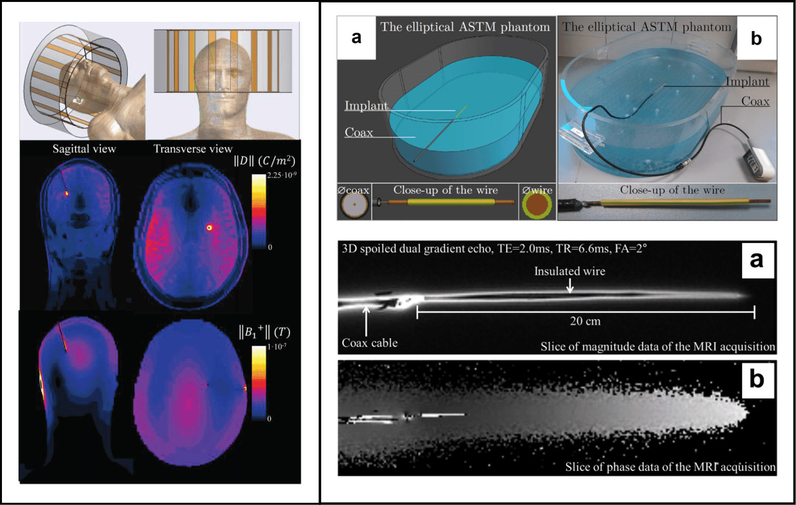
Figure 3: left frame: effect of a DBS implant on the electromagnetic field distribution in the brain. Scattered currents in the implant can lead to electric fields and tissue heating. Right frame: experimental method to characterize the response of an implant to RF radiation by using the implants as an antenna.
Relevant publications
- Van Den Bosch MR, Moerland MA, Lagendijk JJW, Bartels LW, Van Den Berg CAT. New method to monitor RF safety in MRI-guided interventions based on RF induced image artefacts. Med Phys. 2010. https://doi.org/10.1118/1.3298006
- Tokaya JP, Raaijmakers AJE, Luijten PR, Sbrizzi A, van den Berg CAT. MRI-based transfer function determination through the transfer matrix by jointly fitting the incident and scattered B1+ field. Magn Reson Med. 2020. https://doi.org/10.1002/mrm.27974
- Stijnman PRS, Tokaya JP, van Gemert J, et al. Accelerating implant RF safety assessment using a low-rank inverse update method. Magn Reson Med. 2020. https://doi.org/10.1002/mrm.28023
People

Mathijs Kikken

Christina Louka

Mike Eijbersen

Peter Stijnman


Bart Steensma

Nico van den Berg
